

Keywords
Mesenteric vein partial thrombosis
Thrombosis
Mesenteric vein
Hepatopathy
How to Cite
Abstract
Background: Intestinal ischemia has various causes and presentations that can be classified as acute or chronic. They are also subdivided into arterial, venous and non-occlusive causes. Mesenteric venous occlusion predominates in approximately 10% of intestinal ischemia cases. Venous thrombosis are closely related to coagulability problems, as well as other diseases such as antiphospholipid antibody syndrome, polycythemia vera, portal hypertension, or even the use of contraceptives or the state of hypercoagulability in pregnancies.
Case Presentation: A 52-year-old male patient who presents a chronic hepatopathy with portal hypertension, hepatosplenomegaly and with insidious abdominal pain that makes him consult with a gastroenterologist, whom suggests for an abdominal ultrasound. The patient was on a fasting state for examination purposes, in which an increased echogenictity was found in the liver, with a micronodular surface, no nodules or masses, no dilation of intra or extrahepatic bile ducts and with a permeable portal and suprahepatic veins with normal size and appearance. Color-doppler showed a normal flow and thickness. Biliary vesicle without lithiasis. Pancreas with normal morphology and appearance, however inferior and medial to the pancreatic head, there is a hypoechoic tubular, heterogeneous area with irregular edges.
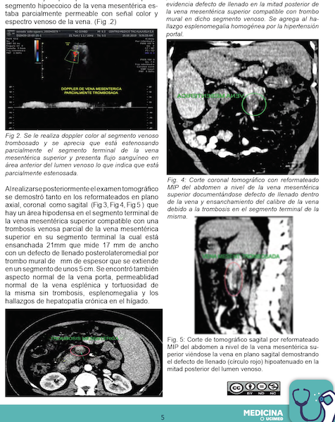
Discussion: The venous circulation can also be compromised on intestine strangulation as observed in volvulus, intussuception or obstruction in the intestinal loop. Acute mesenteric vein thrombosis most commonly occurs when there are pathological conditions such as hypercoagubility states or proliferative neoplasms causing thrombosis of small venous branches with the consequent drastic reduction of venous return and collateral vessel formation. Several imaging methods are currently available for diagnosis, each has its advantages and disadvantages. Among these the conventional high resolution ultrasound, color-doppler ultrasound, multislice computed axial tomography performed with I.V contrast media, which were used in this case can be mentioned. It can also be used as a great contribution the MRI (angiosonance with gadolonium), which in this case was not used, since the diagnosis was clear and due to economic limitations. As another option, a digital angiography of the mesenteric arterial and venous system can be performed, but this procedure is reserved for cases of complicated diagnosis with non-invasive techniques.
Conclusion: Due to the revised information and the catastrophic prognosis of the disease, the greatest efforts should be made to think of this entity in patients with abdominal pain and risk factors and thus exploit further the diagnostic possibilities that can be provided by computed axial tomography, conventional ultrasound and color-doppler ultrasound.

This article is licensed under a Creative Commons Attribution-NonCommercial-NoDerivs 3.0 Unported License. The authors keep the copyright and publication rights in the journal the right of the first publication and this possibility to edit, reproduce, distribute, expose and publicly communicate on the magazine's website. Likewise, it assumes the commitment on any litigation or claim related to the rights of intellectual property, exonerating of responsibility to the Science and Health Magazine of the UCIMED. In addition, you can see how they are published in this journal (eg, Include in an institutional repository or publish it in a book) as long as they clearly indicate the work published for the first time in the magazine Science and Health of the UCIMED.